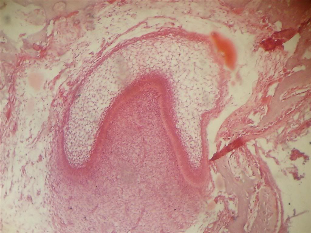
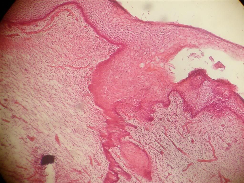
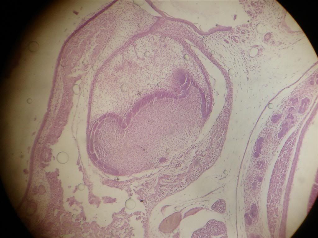
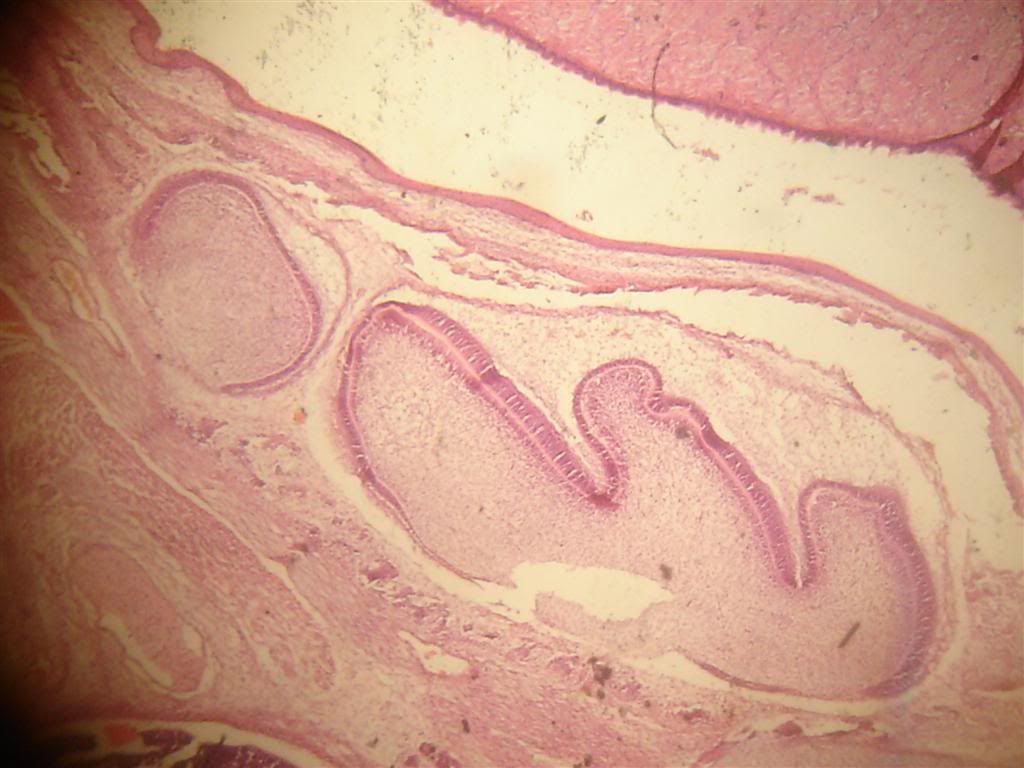
Bismillah. These are photos of some of the jars. The important part here is to get used with the structure so you can easily determine the structure without having to take a long time as the practical exam has more than a dozen question and about 1-2 minutes allowed per question.
Between, it is favorable to borrow the medical student practical book - Mansoura Atlas Part 2. And I personally think the Simplified Practical Anatomy Part 2 also helps a lot.
For those who didn't come to the revision or still blurred about the answer scheme and how the examination works, here it is. (based on last year's exam)
1. There will be multiple microscope in a row. You have a piece of paper, and a doctor to watch over you.
2. Every one will stand infront 1 microscope then the exam start, and you need to see the slide given and answer the question.
3. Answer the question, then within 1-2 minutes, the doctor will instruct to move to another slide (Next!!). Time is crucial, so you should really know every slide with its structure before exam.
4. You may/may not be given a time to check your answer and write your name, group number, etc.
5. Finish.
- The question will be
"Identify the structure shown."
or maybe
"Identify and name the structure within it."
- The answer should be in this scheme:
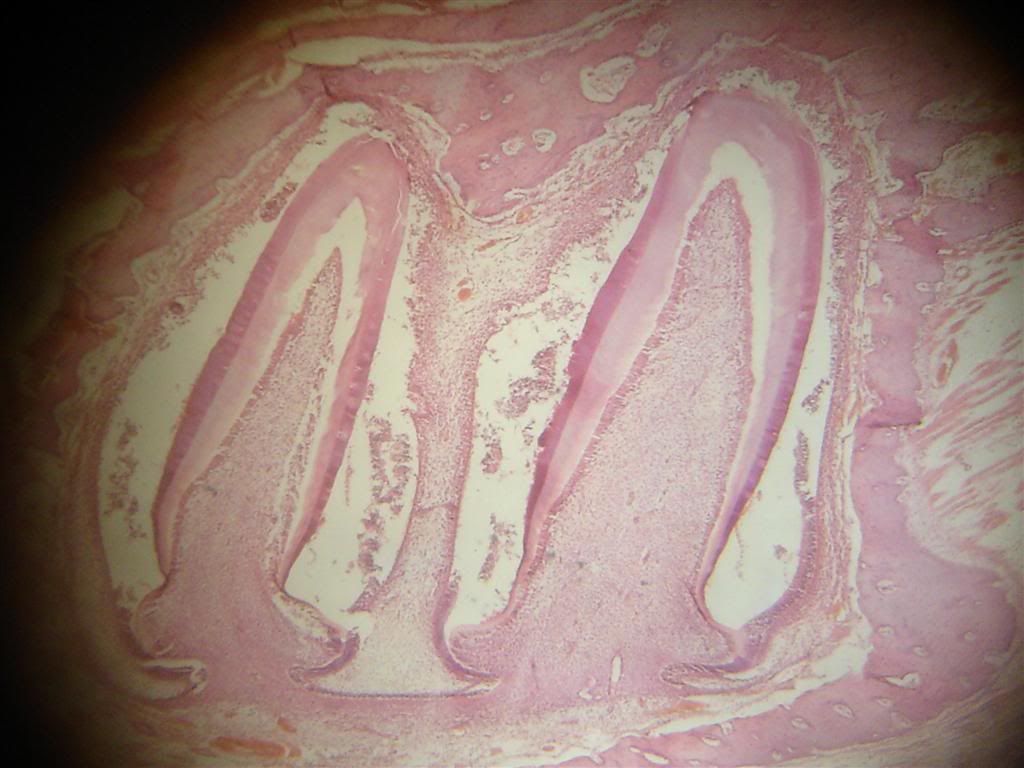
The decalcifiedlongitudinal section using Hx&E (HxE) showing Late Bell Stage of Tooth Development. 1. Ameloblast 2. Outer Enamel Epithelium 3. Inner Enamel Epithelium 4. Stellate Reticulum 5. Stratum Intermedium 6. Dental papilla 7. Odontoblast 8. Predentin
-The main points here are:
1. State wether its decalcified section or ground section. 2. Wether its longitudinal or transverse section. 3. If decalcified slide, state the stain used : Hematoxylin and Eunosin (Hx&E), Silver Stain, Iron Hematoxylin. 4. State the slide is a structure of ... 5. Then give every structure you can see in the slide.
Tips: Sometime, you can see the slide with naked eye and determine what it is. For example, if the microscope's lens pointed to tip of the crown, then you know, it's a structure within the crown (enamel, dentin etc.)
Hope it help a lil bit.
p/s: announcement from mrs Manal :
BERITA BAIK TUK TAHUN1 DENTISTRY. Siapkan dan hantar practical book physio untuk mendapatkan 5 markah carry ke final ganti midtterm exam soalan E.S.R yg sgt buruk markahnya menurut doc physio td. nanti final oral exam physio akan jd 30 markah drp 35markah.sbb dtolak tuk ESR.hantar lps winter break vacation kat department physio.huhu tq doc!sebar2kan. MaNaL: SALAM...BERITA GEMBIRA TUK TAHUN1 DENTIST----->minggu depan selasa 26hb InsyAllah akan ada exam practical oral histologi.pg tadi ada revision tuk slide2 yg akan masuk exam.sapa2 yg xpg revision td lehla ziarah member2 discuss pasal slide tuk kita sama2 dapaT fullmark tuk practical oral histo sbyk 20mark(midterm+final) tuk skema pmarkahan lehla bkunjung ke blog abrar mahmud .Tuk akhawat d alu2 kan tuk dtg ziarah umah sy d syarie samanoudi tuk dptkan slide oral histo ..slide subjek lain pon ade gak,jar anat bla2(promot la pulak ) .moga sama2 kita rebut full mark. sama2 bkongsi ilmu yg ada bia pon sikit kan jd bukit (1+1 =2).insyAllah. p/s : kejayaan milik bersama.. bittaufiq wannajah!
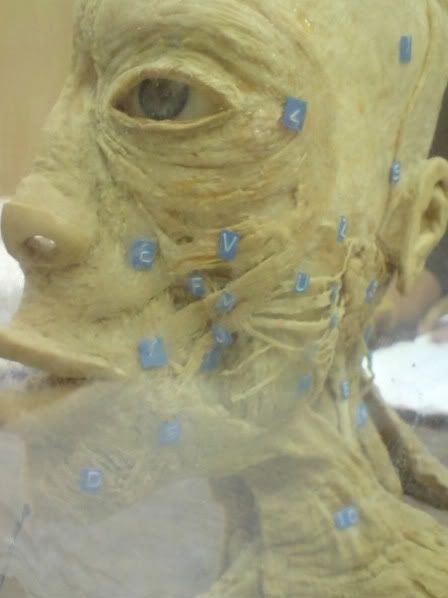
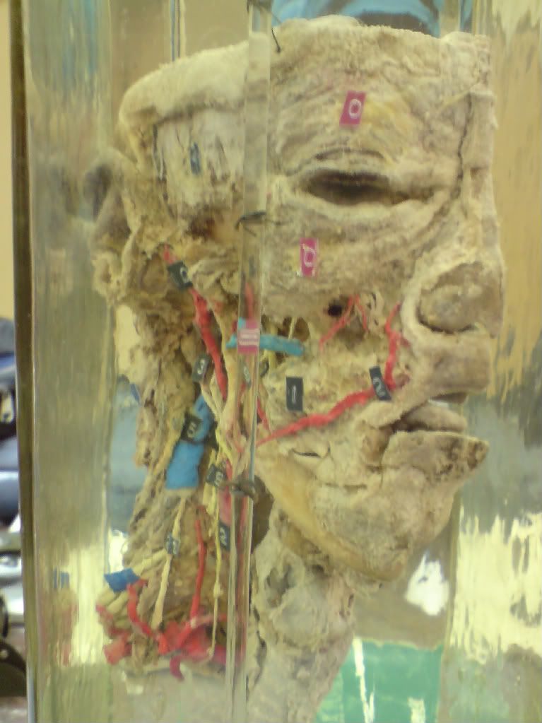
These photos are taken 2 years ago during final revision for final year exam 2007.I don't know if you guys are taking the same things for this year.Hopefully these photos will help you guys out.
P/s: Zoology is one of the hardest subject for pre-dentistry students(Even Egyptian said that!).Try to have a constant and relax study for this subject.








+Ascaris.JPG)