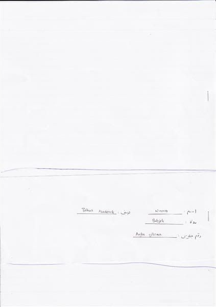
Isi kertas depan (kepilan kecil) dengan maklumat sepatutnya seperti yang diberitahu pengawas.
Mulakan menjawab dari kiri ke kanan (belakang ke depan).
Gariskan pada setiap muka di sebelah kiri dengan garisan vertical.
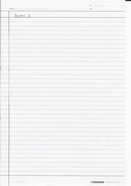
Sehelai kertas (2 muka) untuk satu soalan sahaja. Bila dah habis jawab, gariskan horizontal di bawah jawapan terakhir itu.
Jika jawapan lebih dari satu helai, pada helaian pertama – muka kedua (bawah sekali) buatkan anak panah ke helaian ke dua dan sambung menjawab. Bila selesai menjawab, gariskan garisan horizontal di bawah jawapan. Kemudian mulakan menjawab soalan baru di helaian baru.
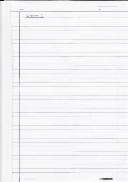
Pada setiap permulaan helaian, tuliskan nombor soalan seperti – “Question 1”
Tgk gambar kalau agak tak faham.
Isi kertas kat depan.
Mulakan dgn tulis nombor question, then garis di kiri page ni.
Di muka 2 helaian yg sama, kalau dah habis jawab (katakan full page), gariskan horizontal di bawah jawapan tu.
Mulakan jawab soalan baru di helaian baru
Kalau sehelai tak cukup, buatkan anak panah di muka 2 helaian itu (bawah).
Then, kalau habis sekerat muka je, gariskan horizontal di bawah jawapan tu.
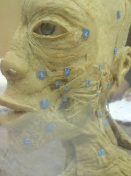
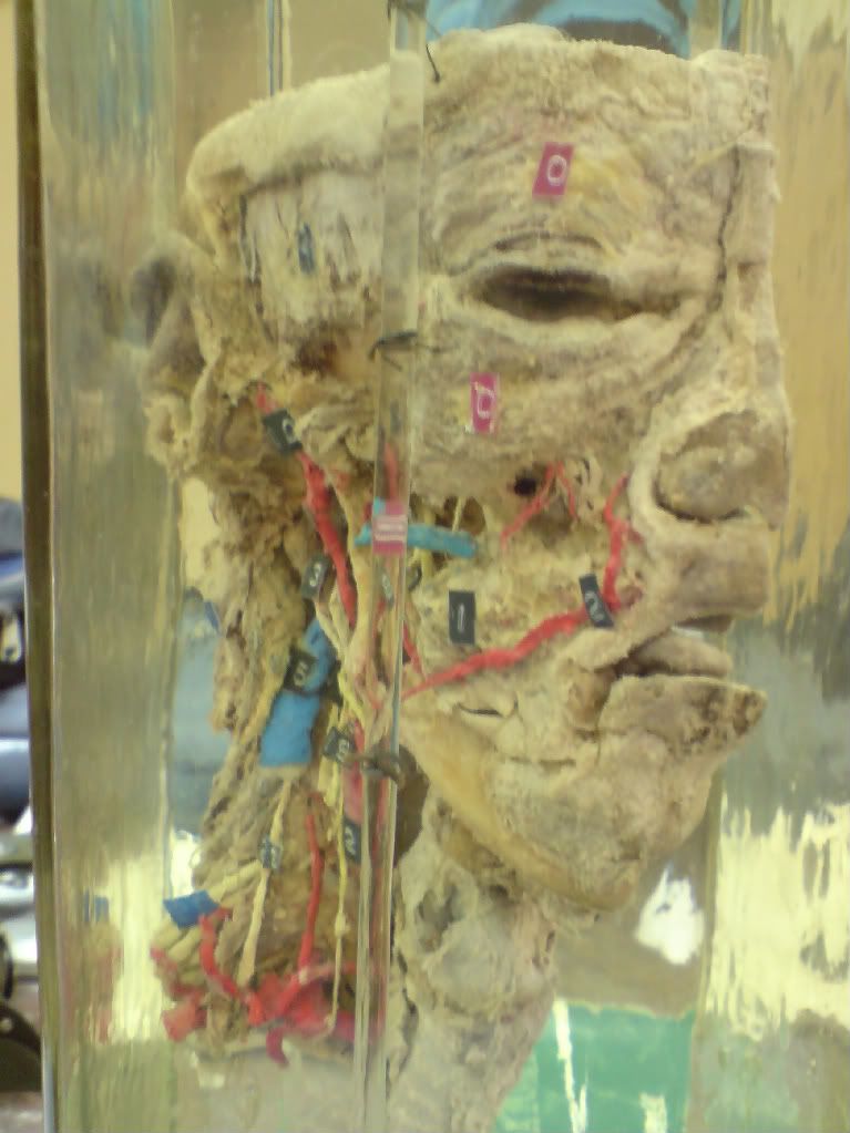
Bismillah. These are photos of some of the jars. The important part here is to get used with the structure so you can easily determine the structure without having to take a long time as the practical exam has more than a dozen question and about 1-2 minutes allowed per question.
Between, it is favorable to borrow the medical student practical book - Mansoura Atlas Part 2. And I personally think the Simplified Practical Anatomy Part 2 also helps a lot.
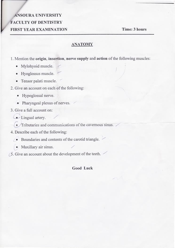
For those who didn't come to the revision or still blurred about the answer scheme and how the examination works, here it is. (based on last year's exam)
1. There will be multiple microscope in a row. You have a piece of paper, and a doctor to watch over you.
2. Every one will stand infront 1 microscope then the exam start, and you need to see the slide given and answer the question.
3. Answer the question, then within 1-2 minutes, the doctor will instruct to move to another slide (Next!!). Time is crucial, so you should really know every slide with its structure before exam.
4. You may/may not be given a time to check your answer and write your name, group number, etc.
5. Finish.
- The question will be
"Identify the structure shown."
or maybe
"Identify and name the structure within it."
- The answer should be in this scheme:
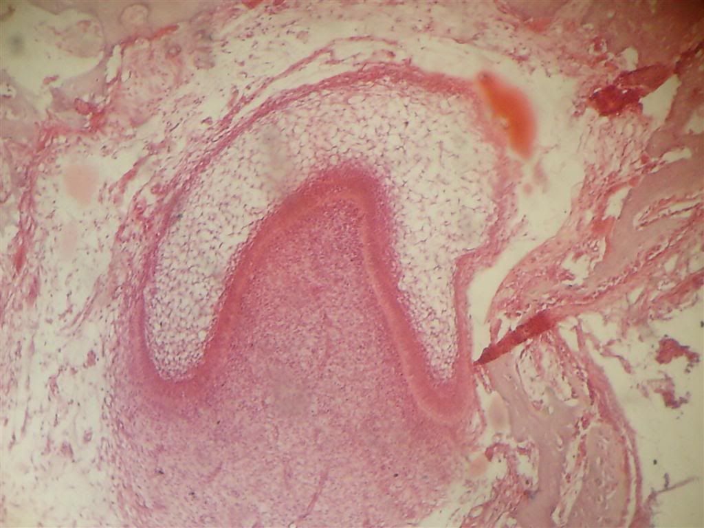
The decalcifiedlongitudinal section using Hx&E (HxE) showing Late Bell Stage of Tooth Development. 1. Ameloblast 2. Outer Enamel Epithelium 3. Inner Enamel Epithelium 4. Stellate Reticulum 5. Stratum Intermedium 6. Dental papilla 7. Odontoblast 8. Predentin
-The main points here are:
1. State wether its decalcified section or ground section. 2. Wether its longitudinal or transverse section. 3. If decalcified slide, state the stain used : Hematoxylin and Eunosin (Hx&E), Silver Stain, Iron Hematoxylin. 4. State the slide is a structure of ... 5. Then give every structure you can see in the slide.
Tips: Sometime, you can see the slide with naked eye and determine what it is. For example, if the microscope's lens pointed to tip of the crown, then you know, it's a structure within the crown (enamel, dentin etc.)
Hope it help a lil bit.
p/s: announcement from mrs Manal :
BERITA BAIK TUK TAHUN1 DENTISTRY. Siapkan dan hantar practical book physio untuk mendapatkan 5 markah carry ke final ganti midtterm exam soalan E.S.R yg sgt buruk markahnya menurut doc physio td. nanti final oral exam physio akan jd 30 markah drp 35markah.sbb dtolak tuk ESR.hantar lps winter break vacation kat department physio.huhu tq doc!sebar2kan. MaNaL: SALAM...BERITA GEMBIRA TUK TAHUN1 DENTIST----->minggu depan selasa 26hb InsyAllah akan ada exam practical oral histologi.pg tadi ada revision tuk slide2 yg akan masuk exam.sapa2 yg xpg revision td lehla ziarah member2 discuss pasal slide tuk kita sama2 dapaT fullmark tuk practical oral histo sbyk 20mark(midterm+final) tuk skema pmarkahan lehla bkunjung ke blog abrar mahmud .Tuk akhawat d alu2 kan tuk dtg ziarah umah sy d syarie samanoudi tuk dptkan slide oral histo ..slide subjek lain pon ade gak,jar anat bla2(promot la pulak ) .moga sama2 kita rebut full mark. sama2 bkongsi ilmu yg ada bia pon sikit kan jd bukit (1+1 =2).insyAllah. p/s : kejayaan milik bersama.. bittaufiq wannajah!
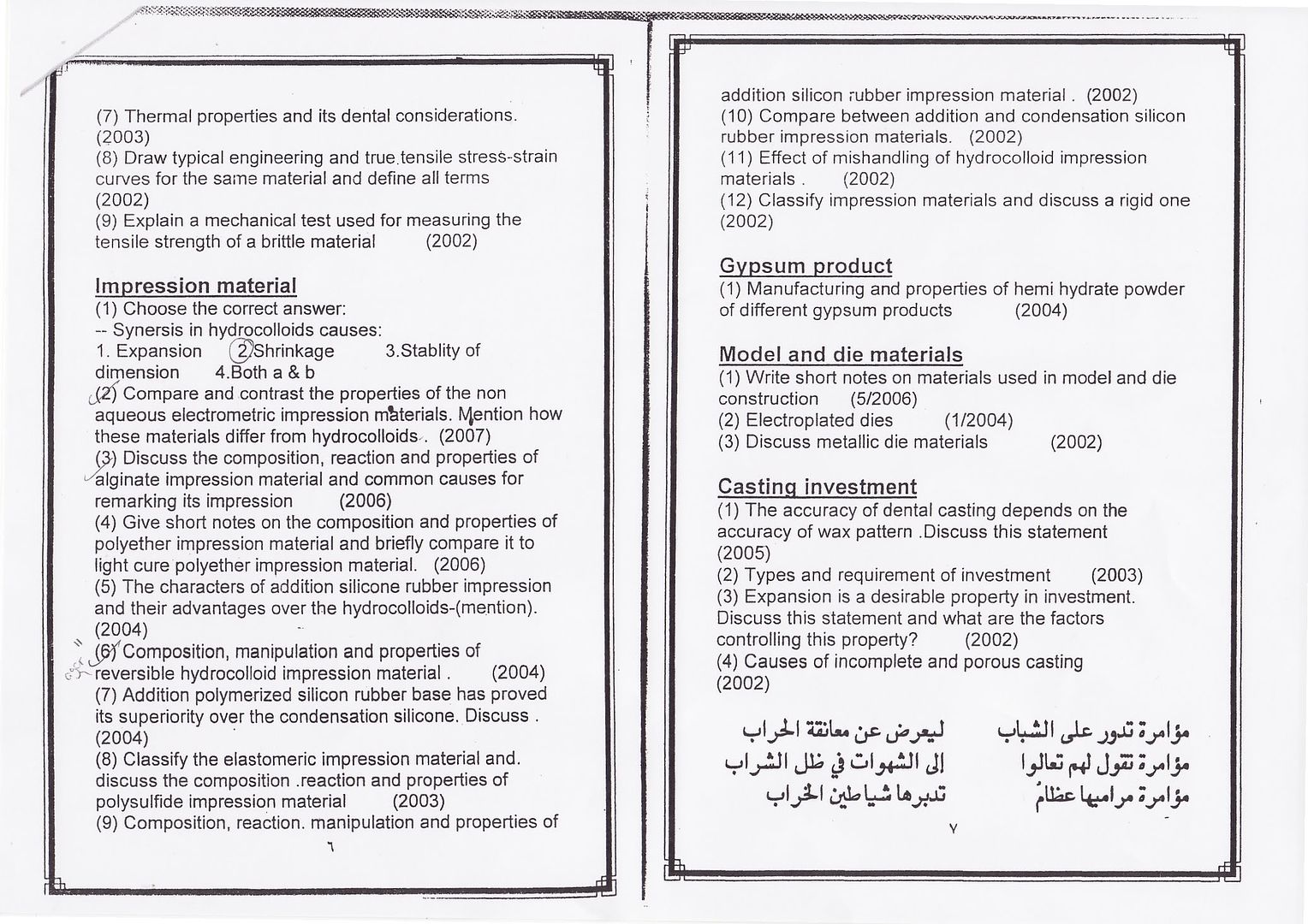
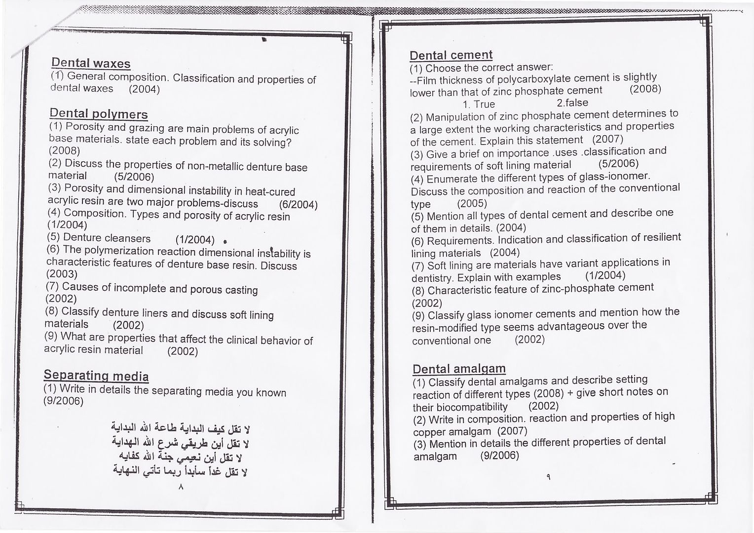
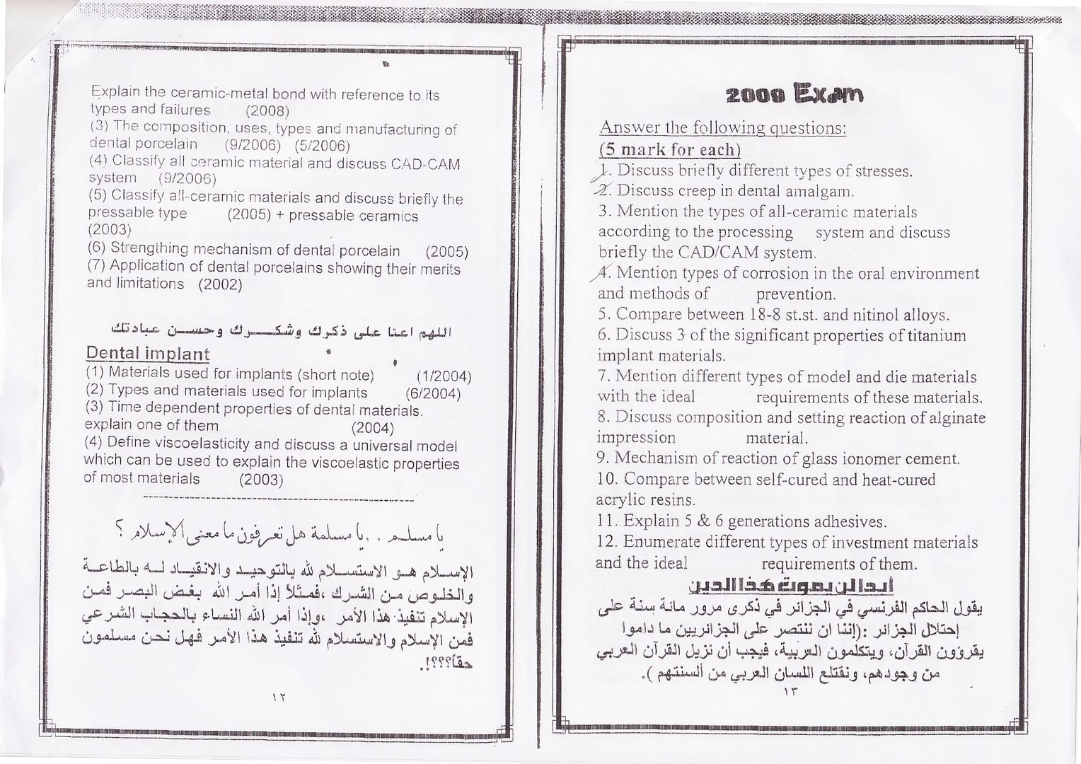
This is the past year question for final exam of Oral Histology by chapters. As for now, it is reasonable to believe that everyone of you have found the best way to master the subject. If not, get going! Lots of way - study in groups or answer those question and submit them to the doc and etc. Let the spirit of exam burns in you.
Extract file tu dengan WinRAR. download kalau takde extractor.
Sample soalan yang ada di dalam file tersebut:
1. MCQ example:
1- What structure is formed in the crown first? a- Enamel. b- Cementum. c- Pulp. d- Dentine.
2- Which of the following induces the dental papilla cells to be differentiated into odontoblasts? a- Stratum intermedium. b- Reduced enamel epithelium. c- Inner enamel epithelium. d- Outer enamel epithelium.
2. True or False
State true or false 1- After destruction of enamel by caries or injury, neither the body nor the dentist can restore the enamel tissue. 2-Enamel is extremely hard because of its high mineral content. 3- The permeability is the property that enables enamel to withstand the mechanical forces applied during tooth functioning.
3. Enumerate
1- Enumerate the stages of odontogenesis 2- Eneumerate the function of inner dental epithelium 3- State the functions of the dental sac.
4. Complete the sentence
1-......................... comprises matrix formation and mineralization of cementum. 2- The ............................ will induce the neighboring cells of the dental papilla to differentiate into odontoblasts. 3-The epithelial root sheath of Hertwig will loss its continuity and its cells become ............................................ 4- The cells entrapped in the mineralized cementum are referred to as ................................and occupy lacunae.
5. Give an account
1- Give an account on four of the following: a) Age changes of the pulp. b) Odontoblastic processes & dentinal tubules. c) Secondary dentine. d) Cellular cementum. e) Odontoblast. f) Cementoblast. g) Accessory root canals
As usual, do it and submit your works to the lecturers. Or discuss with your study group members. Or maybe do it as you think the best way to get the grasp of what its all about. Happy studying.
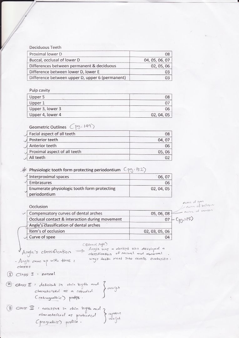
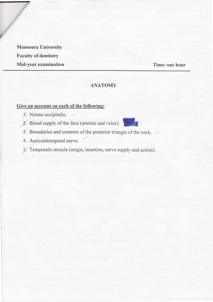
Anatomy: The past year question by chapters Download Ms Word: